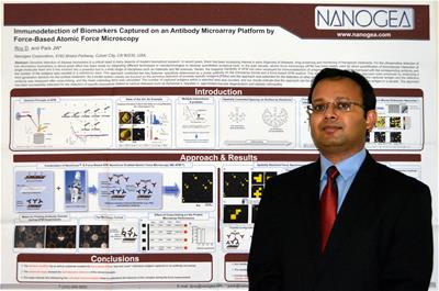
Dhruvajyoti Roy
Sigma Xi member Dhruvajyoti Roy, a senior scientist at Nanogea Corporation in Culver City, California, is on a team that is developing technology for biomedical applications. One way this technology could be used is to improve the way illnesses, such as cancer and Alzheimer's disease, are diagnosed.
For this installment of the Meet Your Fellow Companion series, Heather Thorstensen, Sigma Xi's manager of communications, spoke with Dr. Roy about his work.
Transcript
Heather Thorstensen: Hello and welcome to this Google Hangout from Sigma Xi, The Scientific Research Society. My name is Heather Thorstensen and I’m the manager of communications for Sigma Xi. Sigma Xi connects scientists across disciplines and one of the ways that we do this is through our Meet Your Fellow Companion series, and this is the series where we share the work of our members.
 Today for the series I’m speaking with Dr. Dhruvajyoti Roy of Los Angeles, California. He is a Sigma Xi member and a senior scientist at Nanogea Corporation who is developing an atomic force microscopy, or AFM, based detection technology for biomedical applications. The technology can detect individual molecules and it can detect how single molecules interact. It’s being developed to improve detection of various disease biomarkers. This could improve the way doctors diagnose diseases such as cancer and Alzheimer’s disease, and that’s based on a sample from a patient’s bodily fluids. The technology has broader applications as well. Dr. Roy and his colleagues’ technology could be applicable to improving in vitro fertilization. Welcome to the hangout, Dr. Roy.
Dhruvajyoti Roy: Thank you. Glad to be here.
HT: Let’s start by talking about how this technology is going to be finding these biomarkers. Could you talk about what a biomarker is?
DR: Sure. Actually in general, a biomarker is a measurable indicator of some biological state or disease condition. An ideal biomarker, or disease-related biomarker, obviously has certain characteristics that make it appropriate for checking the severity [of] a particular disease or a particular disease state. There are numerous biomarkers that have been applied for various medical applications and those biomarkers could be protein, could be DNA, RNA, or even a small molecule.
HT: How is the technology that you’re developing going to find these biomarkers, I’m wondering? We have a slide to show for this so I’m going to pull that up while you are explaining it.
DR: Ok. So, here we are focusing on commercializing the industry’s most sensitive single molecule detection platform based on our company’s proprietary NanoConeTM chemistry and NanoCone Enabled Atomic Force Microscopy (NE-AFMTM). In this case of protein biomarkers, our approach combines two key features: the specificity determined by a probe antibody on the microarray platform and the label-free AFM (atomic force microscopy) readout.


Let me explain briefly how the technology works. Typically, to detect a target protein biomarker, a matched pair of antibodies is selected. One of these antibodies is known as the capture antibody and it is spotted on a substrate with a conventional microarray spotter. This is a spatial array of microscopic spots of biological material attached to a solid surface. The other antibody is the detection antibody. That is immobilized on an AFM probe. The capture antibody on a microarray platform specifically captures the target biomarker present in a biological sample from a patient. After capturing, the substrate is washed and then we do a cross-linking step to secure the biomarker on the antibody spot. At this point, the substrate is ready to scan. We employ a force-based AFM approach to detect the captured biomarker directly where the controlled surface architecture allows a single molecular interaction between the detection antibody on an AFM tip and the target biomarker on a substrate. The high-resolution force-based mapping capability of AFM enables us to “see and count” them in a sub-micrometer designated area. The technology can also be utilized for other forms of biomarkers such as DNA, RNA, and small molecules.
HT: And you mentioned one feature of your approach is a label-free AFM readout. Can you explain what that is?
DR: Yes, sure. In general, there are two major strategies for the detection of biomarkers, label-based and label free. The label-based techniques need labelling of either the analyte or the probe molecule with labels such as fluorescent dyes, chemiluminescent tags or radioisotopes. Whereas, label free techniques do not require covalently labelling of the analyte or recognition molecule. Since they avoid the expensive and tedious labeling process and challenging labeling procedures, label-free detection techniques are now attracting significant attention. In this context, force-based AFM is a label-free single molecule technique that directly address the measurement of forces between molecules without any need of labeling.
HT: Could you talk some more about how this technology will actually be used once it’s available on the market?
 DR: Well, the early and accurate detection of the presence of certain biomarkers is really, really important. That actually enables physicians to diagnose cancer and other diseases much earlier. And remarkably, our label-free AFM-based technology can detect a wide range of disease biomarkers and open a really new avenue to an intriguing way of analyzing low-level of biomarkers, particularly in biological samples while it is quite challenging in other existing technologies today. Now, we have successfully utilized this technology for various applications including many forms of cancer, Hepatitis C, and Alzheimer’s disease, among others. Now, the approach has great potential in ultrasensitive biosensing assays that can be applied in biomedical laboratories for routine analysis. We are also focusing to utilize the technology to detect and measure genetic mutations for accurate monitoring of disease prognosis and progressions and treatment [effectiveness,] leading to personalized medicine and healthcare.
HT: When you’re working on this, and you're working towards commercialization, what are you doing day-to-day?
DR: That’s a good question. Actually, my day-to-day activities vary throughout the week. Obviously, the majority of my time is spent in the laboratory developing, evaluating, and obviously integrating the exact state-of-the art technology to improve process development and optimization including experimental design, method, data evaluation, and solve problems related to force-based AFM approaches for bio-analytical applications. In addition, I lead a couple of research projects related to ultra-sensitive detection of biomarkers by our force-based AFM technology in collaboration with major pharmaceutical companies and medical institutes in the United States and around the world.
HT: The technology that you’re developing is not going to be the first to detect biomarkers. How is your technology going to improve [the diagnostics process] that’s already out there?
DR: Most commonly, to detect certain disease-related biomarkers we usually use the conventional enzyme-linked immunosorbent assay, so called ELISA. However, the sensitivity of ELISA is quite limited, and the persistent challenge in this area is the lack of [highly] sensitive readout technologies. In addition, the level of biomarkers for cancers, infectious diseases, and biochemical processes is present at a very low concentration at early stages; so detecting those particular low level concentrations is important and essential for [early] diagnosis of diseases, drug screening, and monitoring therapeutic treatments. The AFM tool is evolving from an imaging instrument to a multifunctional toolbox and it has great potential as an analyzing tool. In a break from the other diagnostic methods, our approach uses that high-resolution AFM-based force spectroscopy mapping to distinguish between the molecules captured on the surface and quantify them. In this aspect, our highly sensitive detection approach can be useful for analyzing biomarkers from very few cells, or even from a single cell so that it can be of great use to detect very low copy number targets.
HT: The part of this that you’re working on is in the nanotechnology sphere and I wonder what other multidisciplinary areas you’re working [with] to get this completed.
DR: Yes, we are working with world-renowned scientific teams and professionals from multidisciplinary areas including biology, chemistry, engineering and medicine. Our technology has tremendous potential for various biomedical applications and obviously, professionals from a range of disciplines working together will help us to comprehensively analyze our data and will address as many of the patient’s need as possible.
HT: What stage would you say that you are in for commercializing this technology?
DR: We have already utilized the technology for detecting various biomarkers related to certain diseases. At this stage, we are optimizing and validating our approach for clinical trials. We are collaborating with doctors, major pharmaceutical and diagnostic companies around the world to clinically validate our technology for wide range of applications. Obviously, we are focusing on commercializing the technology.
HT: What other challenges are you trying to overcome as you’re preparing this technology?
DR: To provide fast, accurate, and reliable and obviously efficient information is always challenging for a diagnostic company. Although, we can detect a wide range of biomarkers unprecedentedly, we are focusing on the speed of our assay. That is really important. We like to complete the detection and analysis of data within two or three hours. For that, we are collaborating with AFM manufacturers, we are working with software specialties to improve our workflow.
HT: Let’s go back to talking about how your technology is going to be used with in vitro fertilization. You mentioned how the technology is going to help doctors select the best-quality embryos. Could you talk about how that’s going to improve the process of in vitro fertilization?
DR: Sure, definitely. Well, one of the major factors that can influence the success of an IVF cycle is the selection of the best embryo for transfer to the uterus. Currently, most clinics routinely transfer multiple embryos in order to be more certain of getting success. However, it more dramatically increases the rate of multiple pregnancy, for example twins, triplets, etc., and we know about the story of “octomom.” So, one of the best solutions to the multiple pregnancy problems is single embryo transfer by finding and selecting the best quality embryo.
HT: Ok, we got through all the questions I wanted to ask you, is there anything else you wanted to say about your work?
DR: I strongly believe that our molecular approach to the diagnosis and detection of disease will also help for a transition to individualized medicine switching from general systemic diagnostic and treatment model to one focused on individual cell structures and cell functions. The result will be a dramatic improvement in both public health and treatment of individual patient.
HT: Ok, Thank you very much.
DR: It’s been a real pleasure to talk to you. Thank you so much.